Lower Extremity Os Foot & Ankle Orthobullets

Pedis X Ray Anatomy Radiology Radiographic Stock Photo 1459919159
This article aims to review the normal anatomy of these bony structures, and to discuss their most common associated pathological conditions.. ossicles. Radiographs confirm the presence of an ossified accessory bone, and fractures are commonly evident on X-rays.. Tandogan R, Gunal I, Arac S. Os vesalianum pedis. J Am Podiatr Med Assoc.

Lower Extremity Os Foot & Ankle Orthobullets
The purpose of this article is to discuss the radiographic assessment of pediatric foot alignment. Clinical scenarios are included to orient the learner to the evaluation of pedi-atric foot alignment. Abnormalities discussed include, but are not limited to, talipes equinovarus (congenital clubfoot), planovalgus, and vertical talus.

Vascular Imaging of the Foot The First Step toward Endovascular
It is useful in diagnosing fractures, soft tissue effusions, joint space abnormalities and localizing foreign bodies in pediatric patients. Patient position the affected leg is externally rotated until the lateral aspect of the foot is resting on the image receptor affected foot is slightly dorsiflexed Technical factors lateral projection
Dorsalis pedis artery Anatomy, branches, supply Kenhub
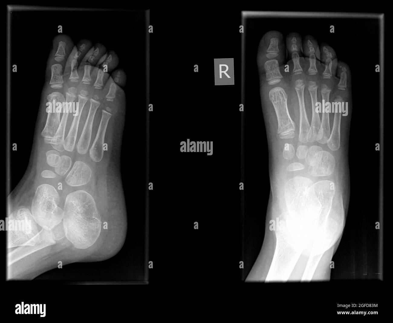
Details of the image 'Normal foot x-rays' Modality: X-ray (Lateral)

Basketball Player With Left Foot Pain
Conventional radiographs are a basic diagnostic tool in pediatric bone imaging (Fig. 1 ). In children with bone and growth disorders, typical indications include the assessment of bone age (e.g., in endocrine diseases), the detection and characterization of physeal abnormalities (e.g., signs of rickets with widening, cupping, and fraying in X.
Osseous injuries of the foot an imaging review. Part 1 the forefoot
Normal chest x ray. Radiological anatomy is where your human anatomy knowledge meets clinical practice. It gathers several non-invasive methods for visualizing the inner body structures. The most frequently used imaging modalities are radiography (X-ray), computed tomography (CT) and magnetic resonance imaging (MRI).X-ray and CT require the use of ionizing radiation while MRI uses a magnetic.
x ray image of fractures and dislocation of phalanx proximal pedis foto
Indications This projection is useful in diagnosing fractures; particularly 5 th metatarsal fractures, soft tissue effusions, joint space abnormalities and localizing foreign bodies in pediatric patients. Patient position the patient is supine with the affected knee flexed plantar aspect of the affected foot resting on the image receptor
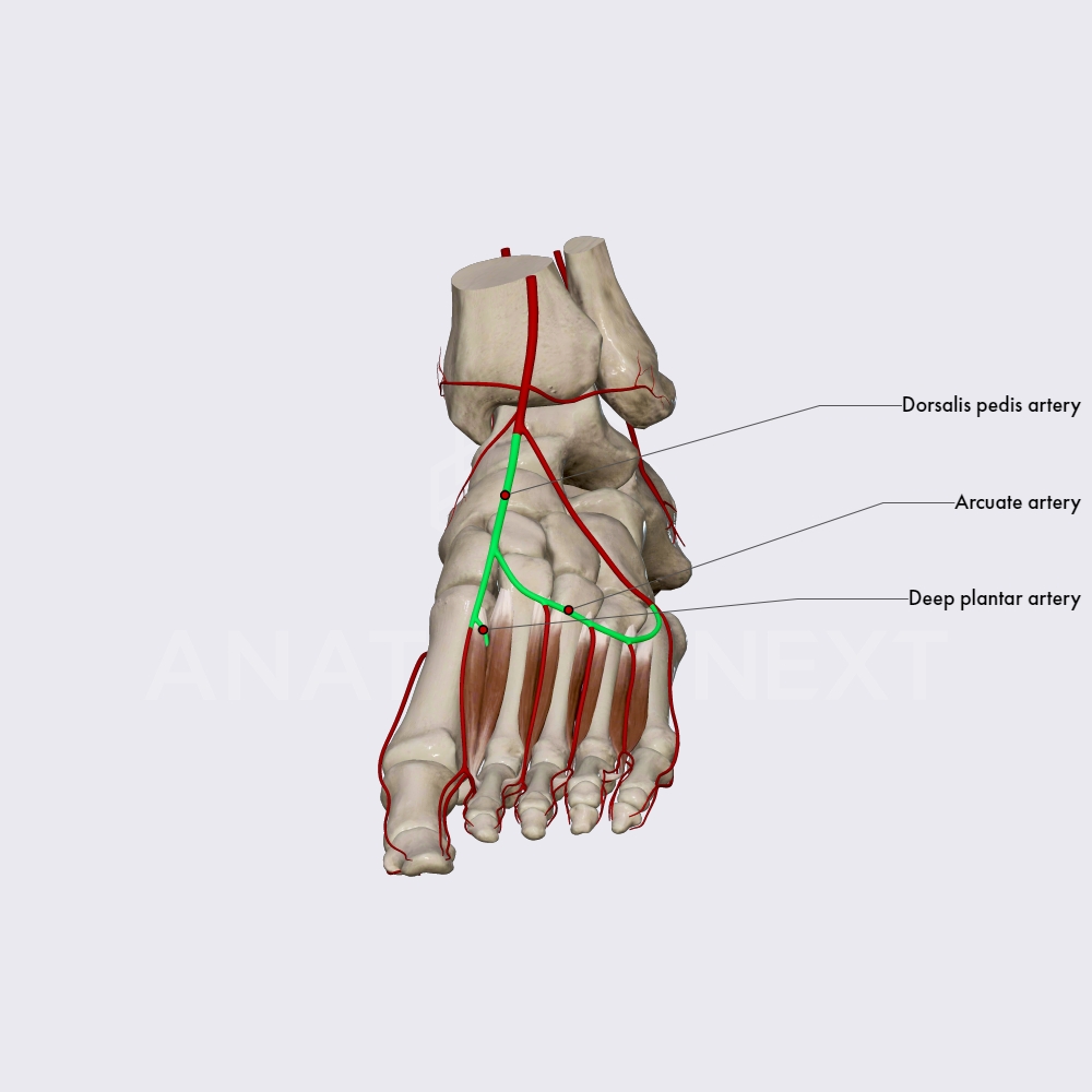
Dorsalis pedis and arcuate arteries Arteries of the lower limb
X-rays use a form of radiation called electromagnetic waves. These waves create a picture of the inner structure (anatomy) of your body. X-rays were first discovered in 1895, making them the oldest type of medical imaging used. The first X-ray took a picture of human tissue in 1896. X-rays are also the most frequently used type of imaging.

Pedis X Ray Anatomy Radiology Radiographic 스톡 사진 1459919177 Shutterstock
Os vesalianum pedis is a rare accessory foot ossicle. It is usually asymptomatic, however, it can be an infrequent cause of lateral foot pain. We present the case of a 19-year-old healthy male.

Pedis X Ray Anatomy Radiology Radiographic Stock Photo 1459919165
Cobey described a posterior view of the foot, modifying the Harris-Beath calcaneal axial view such that distortion was minimized (by directing the x-ray beam perpendicular to the image receptor). The patient stood on a radiolucent platform, and a 14 × 17-inch image receptor was positioned anterior to the patient and extending partially below the platform such that it was tilted 15-20° from.
Podiatrist in Lehi Hallux Rigidus in Lehi Arches Foot and Ankle Clinic
It is useful in diagnosing fractures, soft tissue effusions, joint space abnormalities and localizing foreign bodies in pediatric patients. Patient position the patient is supine with the affected knee flexed plantar aspect of affected foot resting on the image receptor Technical factors anteroposterior projection centering point
Radiografía del pie fotografías e imágenes de alta resolución Alamy
Introduction Infants and children undergo imaging studies to evaluate a wide variety of congenital and acquired disorders. The first imaging study is conventional radiographs; sometimes they are performed with the need for additional imaging which is guided by the plain film findings and clinical presentation.

Стоковая фотография 1459919189 Pedis X Ray Anatomy Radiology
This is a repository of radiograph examples (X-rays) of the pediatric (children) skeleton by age, from birth to 15 years. Ages are approximate (generally, at most +/- 1-2 months, but mostly within + / - 15 days - unless stated otherwise). Male and female subjects are intermixed. These normal bone xrays are NOT intended as bone-age references!

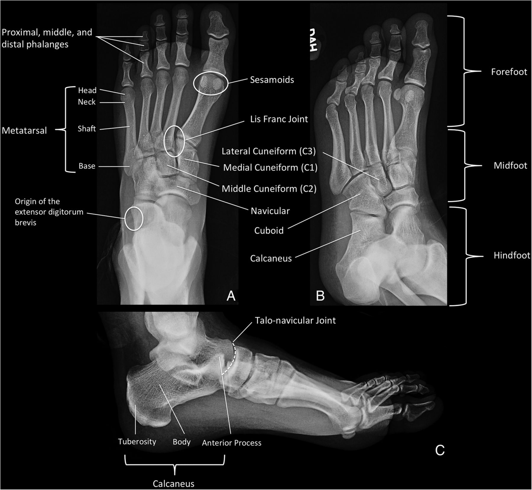
Foot Radiographic Anatomy wikiRadiography Radiology student
First cuneiform (medial) Medial sesamoid. Lateral sesamoid. Shaft of second metatarsal. Neck of third metatarsal. Head of fourth metatarsal. Metatarsophalangeal joint. Proximal phalanx. Middle phalanx.
/Dorsalpedisartery-23c28539f764403ca9254e32551049ce.jpg)
Dorsalis Pedis Artery Anatomy, Function, and Significance
Adult acquired flatfoot deformity (AAFD) is a common disorder that typically affects middle-aged and elderly women, resulting in foot pain, malalignment, and loss of function. The disorder is initiated most commonly by degeneration of the posterior tibialis tendon (PTT), which normally functions to maintain the talonavicular joint at the apex of the three arches of the foot. PTT degeneration.
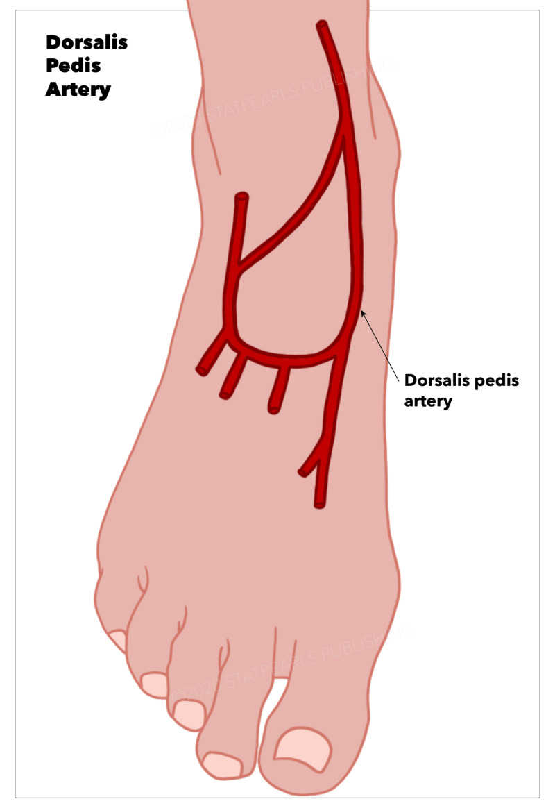
Anatomy, Bony Pelvis and Lower Limb Foot Dorsalis Pedis Artery
Musculotendinous Extensor tendons attach on the dorsal aspect of the foot. In the great toe, the distal phalanx receives extensor hallucis longus whereas the proximal phalanx receives extensor hallucis brevis (part of extensor digitorum brevis ). An extensor expansion also exists, formed from extensor digitorum, the lumbricals and interossei.
